← MAITRI Lab
 ChakshuRakshak AI
ChakshuRakshak AI
ChakshuRakshak AIThree analyses in one platform: an ICDR diabetic retinopathy grade with vessel and lesion maps, a diabetic macular edema grade with a hard-exudate map, and a beta screen for other retinal diseases. Upload one or many fundus photographs and download a printable report for each analysis. Built on the lab's fractional enhancement and vascular-geometry research.
A screening aid for ophthalmologists and diabetic eye-screening programmes. Pick the analysis you need; each runs its own model on the images you give it. Results are always reviewed by a qualified ophthalmologist.
A five-stage diabetic retinopathy grade on the ICDR 0–4 scale (No DR to PDR) with a confidence score and referral advice.
Grades each image as no DME, or hard exudates away from or near the fovea, with the probability of each grade.
A research preview that looks past diabetic retinopathy for conditions that can be mistaken for it.
The same workflow for all three analyses, from upload to report.
Upload both eyes or a whole set of fundus photographs. Results open with a summary table and a full panel for every image.
Add patient details and download a printable PDF report for DR, DME or the multi-disease screen, with the images and findings.
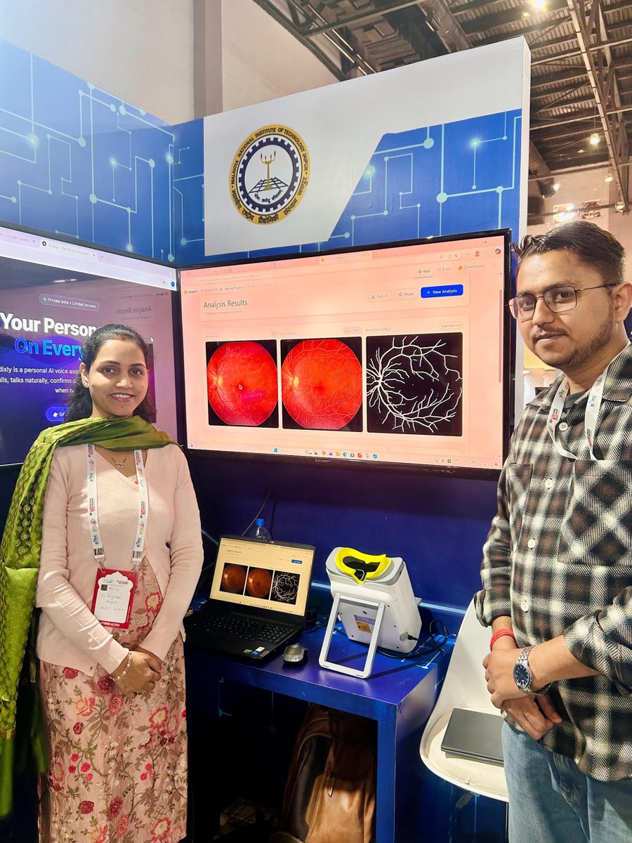
Every result shows the overlay it rests on, such as vessels, lesions or exudates, next to the original photograph.
Run any analysis as a guest. Sign in to keep your DR reports in My Reports.
A web app that runs in the browser on desktops, tablets and phones, with nothing to install.
Built on the lab's fractional-order image enhancement and retinal vessel research, published in peer-reviewed journals.
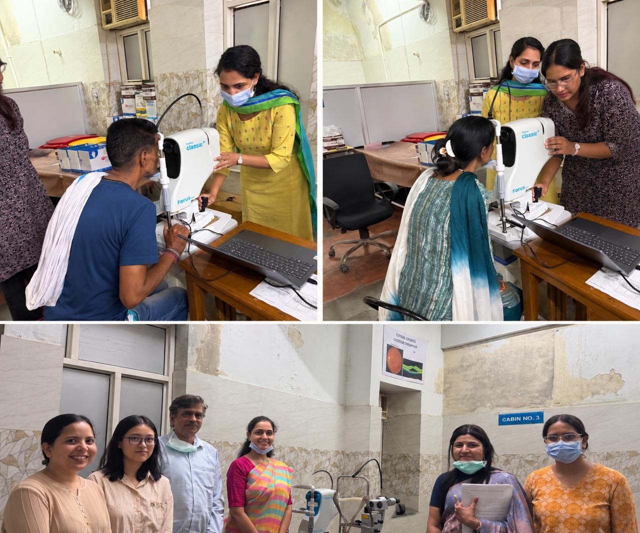
The Directorate of Medical & Health Services, Government of Rajasthan (State Health Society, NCO) approved the pilot implementation and clinical validation of ChakshuRakshak AI for screening diabetic retinopathy and diabetic macular edema, in collaboration with the Department of Medical, Health & Family Welfare.
Weekly progress reports go to the State Directorate. Final diagnosis and treatment stay with qualified ophthalmologists; the AI assists screening.
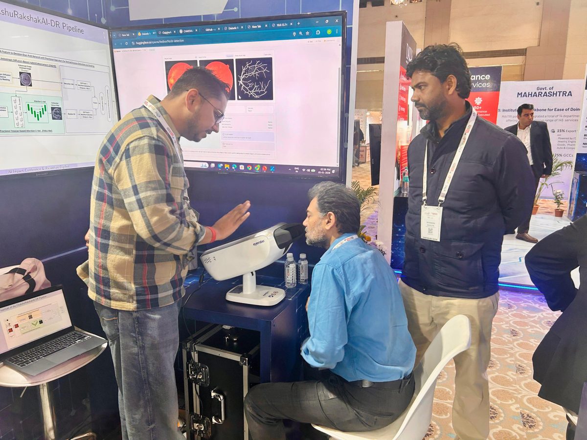
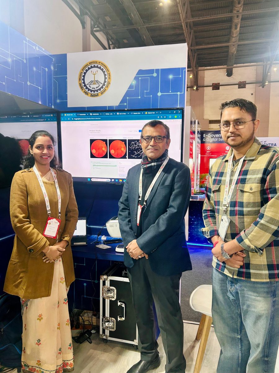
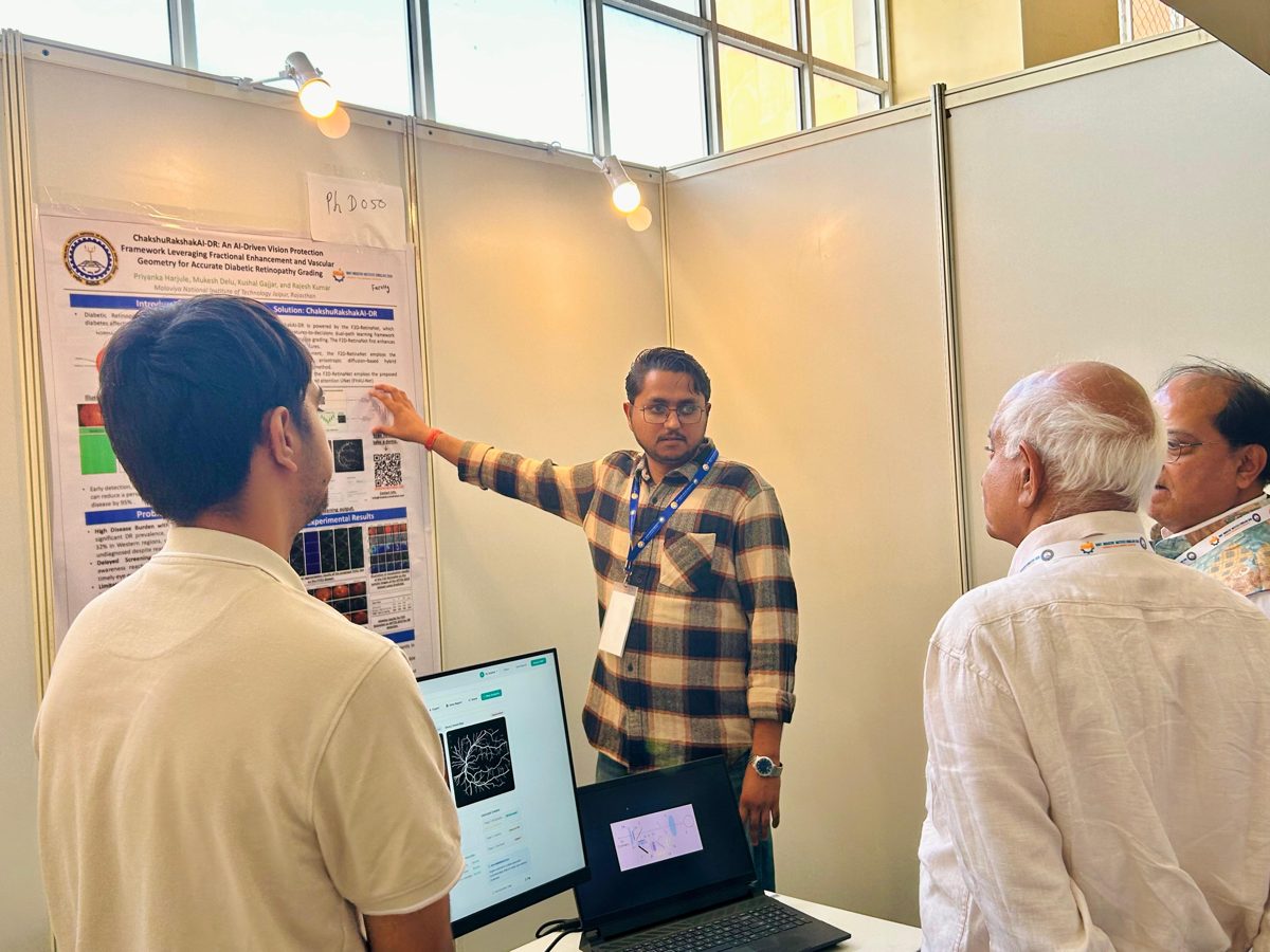
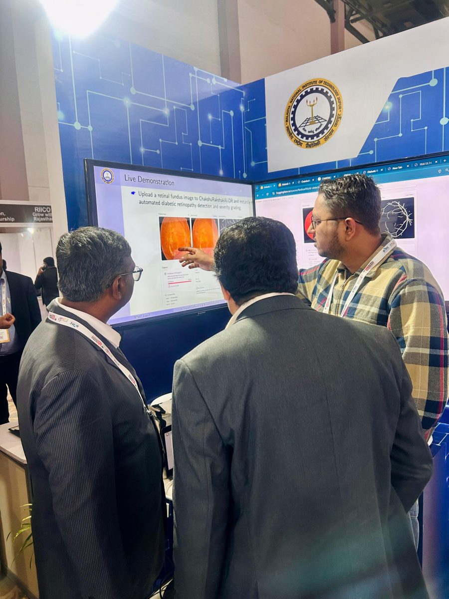
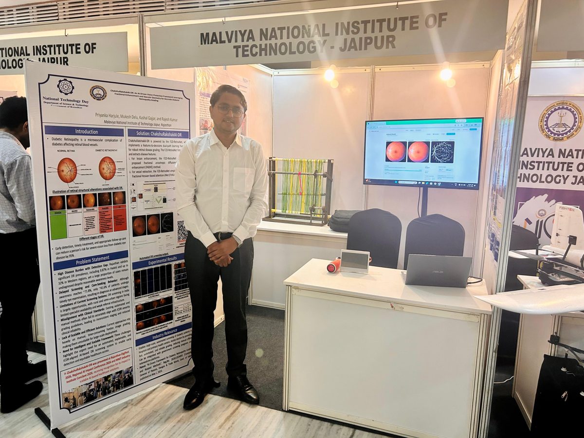
ChakshuRakshak AI was showcased at four state and national events in 2026 and recognised for its impact as a scalable AI healthcare solution.
Papers and the patent that ChakshuRakshak AI is built on.
Priyanka Harjule, Mukesh Delu, Rajesh Kumar, Kushal Gajjar · Reg. No. 202611024093 · Controller General of Patents, Designs & Trade Marks, Govt. of India
Hospitals, screening programmes and researchers interested in ChakshuRakshak AI can try the demo or write to us at info@chakshurakshakai.com or priyanka.maths@mnit.ac.in.